The Imaging Laboratory is in Room 6245 on the 6th floor of the Brehm Center for Diabetes Research (see maps below). The Core consists of a suite of interior rooms (6245, 6245A and 6245B) and each microscope is housed in an individual room to maximize imaging of fluorescent signals. The main area of the core consists of a laboratory with 2 benches for specimen preparation, a fume hood and space for two computer workstations and printers. In addition, the Core has access to shared space including autoclave and dish washing facilities. Tissue culture incubators are present for short-term storage of cells under a controlled environment.
The Core’s aims are to provide service, access to specialized state-of-the-art instrumentation, education and training in morphological techniques.
Map to Brehm Tower
Interior Map of Brehm Tower 6th Floor
Our services and equipment are currently run through MiCORES. MiCORES is an online core management system from Agilent Technologies and iLab Solutions designed to streamline the process of requesting and billing for core services.
Confocal Microscopes
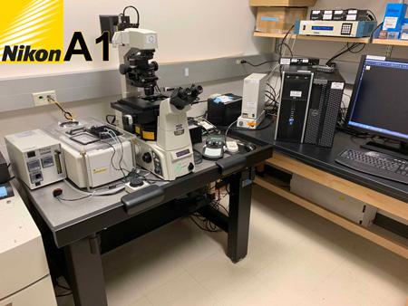
Nikon A1 Confocal Microscope
Nikon A1 Reservation Calendar + LOGIN
Quick view Nikon A1 Calendar no Login
- Nikon TiE Inverted Microscope
- Designed for Live Cell Imaging
- Prior motorized stage for tiling large areas and multi-point mark and find
- Nikon’s Perfect Focus System (PFS) to maintain focus on sample by compensating for thermal drifts
- Tokai Hit environmental chamber to regulate temperature and CO2
- Filter based system to direct wavelengths to the PhotoMultipler Tube detectors for confocal microscopy
- Cy-5 filter set for visualizing far red signals in the oculars/eyepieces before imaging with the laser
- Located in Room 6245B Brehm Center
Wide field Components
- Highly sensitive (95% quantum efficient) Photometrics Prime 95B cMOS monochrome camera for widefield microscopy
- Sutter filters wheels for standard blue, green and red fluorescent signals and CFP and YFP for FRET (Förster resonance energy transfer)
Strengths of the Nikon A1 Confocal Microscope
- Designed for Live Cell Imaging
- Prior motorized stage for tiling large areas and multi-point mark and find.
- Nikon’s Perfect Focus System (PFS) to maintain focus on sample by compensating for thermal drifts.
- Tokai Hit environmental chamber to regulate temperature and CO2.
- Filter based system to direct wavelengths to the PMT detectors.
- Cy-5 filter set for visualizing far red signals in the oculars/eyepieces before imaging with the laser.
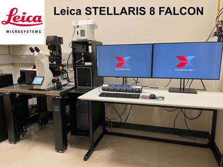
Leica Stellaris 8 FALCON Confocal Microscope
Leica Stellaris 8 Reservation Calendar + LOGIN
Quick view Leica Stellaris 8 Calendar no Login
- Leica DMi 8 Inverted Microscope
- 8 kHz Tandem Resonant Scanner
- White Light Laser -440 nm -790 nm and 405 nm Laser Diode
- AOBS (Acousto Optical Beam Splitter). Tunable crystal that allows for precise laser line excitation for up to 8 lines simultaneously
- Spectral Detectors for variable emission wavelength selection and lambda (wavelength) scanning for spectral unmixing
- 5 Power HyD Detectors (2 x HyD S, 2 x HyD X, HyD R-out to 850 nm)
- FALCON fluorescence lifetime microscopy (FLIM, includes Phasors and FCS)
- Scanning Stage SuperZ for precise control over Z scans, tiling of large areas and multi-point mark and find
- Closed Loop Focus with Adaptive Focus Control
- ToKai Hit Stage Top Incubator
- Leica LIGHTNING Super Resolution (Deconvolution)
- Software Wizards for programming specialized techniques like FRET, FRAP and photoactivation
- Located in Room 6245A Brehm Center
Publications that include data collected with the Leica STELLARIS 8 system must acknowledge support of the S10 shared instrument as follows:
“This work used a Leica STELLARIS 8 FALCON Confocal Microscopy System, funded by a National Institutes of Health SIG grant NIH S10OD28612-01-A1 and available through the Microscopy and Image Analysis Core of the Michigan Diabetes Research Center, funded by National Institutes of Health Grant P30 DK-20572 from the National Institute of Diabetes and Digestive and Kidney Diseases.”
...or...
“This work used a Leica STELLARIS 8 FALCON Confocal Microscopy System that was purchased with funds from a National Institutes of Health SIG grant NIH S10OD28612-01-A1.”
...or...
“The authors would like to acknowledge the NIH S10 Shared Instrumentation Grant NIH S10OD28612-01-A1 for supporting this work.”
Strengths of the Leica Stellaris 8 FALCON Confocal Microscope
- 8 kHz Tandem Resonant Scanner for rapid scanning for live cell imaging or stitching large areas of tissue.
- LIGHTNING deconvolution acquisition and post processing software.
- FALCON fluorescence lifetime microscopy (FLIM, includes Phasorplotsand FCS)
- Tokai Hit environmental chamber to regulate temperature and CO2
- Software Wizards for programming specialized techniques like FRET, FRAP and photoactivation.
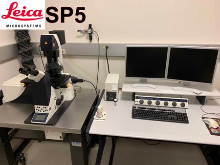
Leica SP5 Confocal TCS Microscope
Leica SP5 Reservation Calendar + LOGIN
Quick view Leica SP5 Calendar no Login
- Leica DMI 6000 Inverted Microscope
- AOBS (Acousto Optical Beam Splitter). Tunable crystal that allows for precise laser line excitation for up to 8 lines simultaneously
- Spectral Detectors for variable emission wavelength selection and lambda (wavelength) scanning for spectral unmixing
- Scanning Stage SuperZ for precise control over Z scans, tiling of large areas and multi-point mark and find
- Software Wizards for programming specialized techniques like FRET, FRAP and photoactivation
- Located in Room 7054 Brehm Center
Strengths of the Leica SP5 Confocal TCS Microscope
- AOBS (Acousto Optical Beam Splitter). Tunable crystal that allows for precise laser line excitation for up to 8 lines simultaneously.
- Spectral Detectors.
- Allows for variable emission wavelength selection.
- Lambda (wavelength) scanning for spectral unmixing.
- Scanning Stage SuperZ. Allows for precise control over Z scans, tiling of large areas and multi-point mark and find.
- Software Wizards for programming specialized techniques like FRET, FRAP and photoactivation.
Epi-Fluorescence Microscope
Nikon TiE Widefield Imaging System

Nikon A1 Reservation Calendar + LOGIN
Quick view Nikon A1 Calendar no Login
- Highly sensitive (95% quantum efficient) Photometrics Prime 95B cMOS monochrome camera for widefield microscopy
- Sutter filters wheels for standard blue, green and red fluorescent signals and CFP and YFP for FRET (Förster resonance energy transfer)
- Located in Room 6245B Brehm Center
High Resolution Image Processing Workstations and Supporting Software
The Core has four high-end imaging workstations based on Windows 64-bit operating systems.
- Leica Application Suite X: Lightning Deconvolution, 3D Visualization, Fluorescence Lifetime Imaging Microscopy (FLIM) & phasor plots, and LIGHTNING deconvolution
- Nikon Elements
- MetaMorph version 7.10 Offline Analysis
- Imaris version 9.5: 3D and 4D Imaging
- Aivia version 12.1 - Artificial Intelligence - machine and deep learning
- AutoQuant X version X3 : 2D/3D deconvolution
- MATLAB version R2022a
- GraphPad Prism version 9.3 (Graphing & Statistical Analysis)
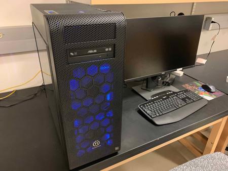
PC Workstation #1: Windows 10 64-bit Operating System
Windows PC #1 Reservation Calendar + LOGIN
Quick view Windows PC #1 Calendar no Login
Specs
- AMD 16-Core Thread ripper 3.4 GHz processor, 128 GB memory, NVIDIA Quadro P4000 8 GB graphics card, 500 and 950 GB solid state hard drives, 6 TB data drive, dual 27” Dell UP2716D 2560x1440 monitors
- Located in Room 6245 Brehm Center
Image Analysis Software
- 3D Imaris, AutoQuant deconvuloution, 3D Volocity, MATLAB, ImageJ, Offline Nikon Elements, Offline Leica Application Suite (LAS X) that includes 3D Visualization, Fluorescence Lifetime Imaging Microscopy (FLIM) & phasor plots, and LIGHTNING deconvolution, ImageJ version 1.46h: Open Source Analysis Software, Fiji version ImageJA 1.45b: Open Source Analysis Software
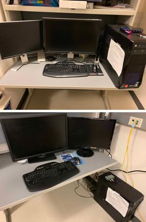
PC Workstation #2: Windows 7, 64-bit Operating System
Windows PC #2 Reservation Calendar + LOGIN
Quick view Windows PC #2 Calendar no Login
Specs
- Intel i7 3.4 GHz 4-core processor, 16 GBs of RAM, Radeon HD 4650 1 GB video card and 128 GB solid state hard drive running Windows 7 32-bit or 64-bit OS, 2 TB secondary hard drive for data storage, 24" Dell UltraSharp U2408WFP 1920x1200 and 19" Dell 1907FP 1280x1024 monitors
- Located in Room 6245 Brehm Center
Image Analysis Software
- MetaMorph version 7.8.6, ImageJ version 1.51k, Fiji version ImageJA 1.5k, Microsoft Office 2016: Word, Excel, PowerPoint, Adobe Creative Suites 6 Web Design Premium: Acrobat X, PhotoShop CS5, Illustrator CS5, Dreamweaver CS5, VirtualDub 64: Video Editing Software
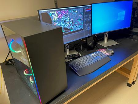
PC Workstation #4: Windows 10 64-bit Operating System HITS FLEX PC
Windows PC #4 Reservation Calendar + LOGIN
Quick view Windows PC #4 Calendar no Login
Specs
- Intel Xeon 6-Core 3.8 GHz processor, 128 GB memory, NVIDIA GeForce RTX 11 GB GDDR6 graphics card, 2 TB solid state hard drives, 6 TB 7200 rpm data hard drive, two 27” Dell U2723QE 3840x2160 monitors
- Located in Room 6245 Brehm Center
Image Analysis Software
- AIVIA Artificial Intelligence-guided Software, MATLAB, Office 365
Additional High Resolution Image Processing Workstations
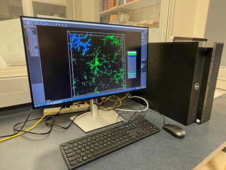
VRC Dell Precision 5820 Offline Image Analysis: Windows 10, 64-bit Operating System, HITS FLEX PC
VRC Dell Precision 5820 Offline Image Analysis Reservation Calendar + LOGIN
Quick view VRC Dell Precision 5820 Offline Image Analysis no Login
Specs
- Intel Xeon 10-Core W-2255 3.7 GHz processor, 128 GB memory, Nvidia RTX A4000 16 GB 4 DP graphics card, 500 GB and 2 TB GB M.2 PCIe NVMe Class 40 solid state hard drives, 27” Dell UP2723QE 3840 x 2160 monitor
- Located in Room 7054 Brehm Center
Image Analysis Software
- 3D Imaris, MATLAB, Free Leica Application Suite (LAS X)
Cryosection Service
Members of the MDRC will be provided access to cryosection services on a fee-for-service basis. Two Leica cryostats for laboratory are Located on the 7th floor of the Brehm Research tower. One cryostat is the CM3050S, which uses disposable blades and incorporates a cooled stage along with the refrigerated cabinet. The second Leica is a CM3050, which utilizes steel knives, without the freezing stage. Both can be used to section fixed or unfixed tissue from 5 to 60 microns, at temperatures from 0 to -35 degrees centigrade. Training on use of the cryostat and access to the systems are provided to MDRC members through the Morphology and Imaging Core of the Vision Research Center.
Other Specialized Equipment on Medical Campus
The core can advise and, in some cases, assist investigators who wish to use imaging instrumentation outside the core. Investigators interested in these services should make special arrangements with Dr. Stephen Lentz (e-mail: lentzs@umich.edu (link sends e-mail) or Telephone: 734-647-8233) for more information.